Home » Unlabelled » Cryptosporidium Oocyst Under Microscope / Cryptosporidium apodemi sp. n. oocysts visualized in ... : In sucrose preparations, the oocysts float just under the coverslip on the slide.
Cryptosporidium Oocyst Under Microscope / Cryptosporidium apodemi sp. n. oocysts visualized in ... : In sucrose preparations, the oocysts float just under the coverslip on the slide.. Food samples are rarely tested for cryptosporidium oocysts or giardia cysts. Cryptosporidium is a genus of protozoan pathogens which is categorized under the phylum the pattern of cryptosporidium life cycle fits well with that of other intestinal homogeneous coccidian a recombinant cryptosporidium parvum oocyst surface protein (rcp15/60) vaccine has produced an. Some people may confuse cryptosporidiosis for cryptococcus, as both sometimes go under the name crypto. cryptococcus is a type of invasive fungus that can cause cryptococcosis. Cryptosporidium is a genus of parasites which has become a rising concern due to its presence in. Epa calls for removal or inactivation of 99.9% of cryptosporidium oocysts in drinking water.
The fourth one either left the oocyst later arrowood mj, sterling cr (1987) isolation of cryptosporidium oocysts and sporozoites using discontinuous sucrose and isopycnic percoll gradients. Cryptosporidium is a genus of protozoan pathogens which is categorized under the phylum the pattern of cryptosporidium life cycle fits well with that of other intestinal homogeneous coccidian a recombinant cryptosporidium parvum oocyst surface protein (rcp15/60) vaccine has produced an. Cryptosporidiosis is caused by protozoan parasites of the genus cryptosporidium (family cryptosporidiidae, order eucoccidiorida, subclass. After cryptosporidium oocysts are ingested, they excyst in the gastrointestinal tract and release sporozoites, which parasitize gastrointestinal epithelial cells. Unstained oocysts are very difficult to see with an ordinary light microscope.
Get Free Stock Photos of Cryptosporidium parvum Protozoa ... from freerangestock.com After cryptosporidium oocysts are ingested, they excyst in the gastrointestinal tract and release sporozoites, which parasitize gastrointestinal epithelial cells. Cryptosporidiosis is caused by protozoan parasites of the genus cryptosporidium (family cryptosporidiidae, order eucoccidiorida, subclass. The fourth one either left the oocyst later arrowood mj, sterling cr (1987) isolation of cryptosporidium oocysts and sporozoites using discontinuous sucrose and isopycnic percoll gradients. Light microscope observations confirmed that relatively often only three sporozoites were released; Bioaccumulation and elimination of cryptosporidium parvum oocysts in experimentally exposed eastern oysters. It spreads through food and water. Unstained oocysts are very difficult to see with an ordinary light microscope. Oocysts (pink arrows) in wet mount.
Survival of cryptosporidium parvum oocysts under various environmental pressures.
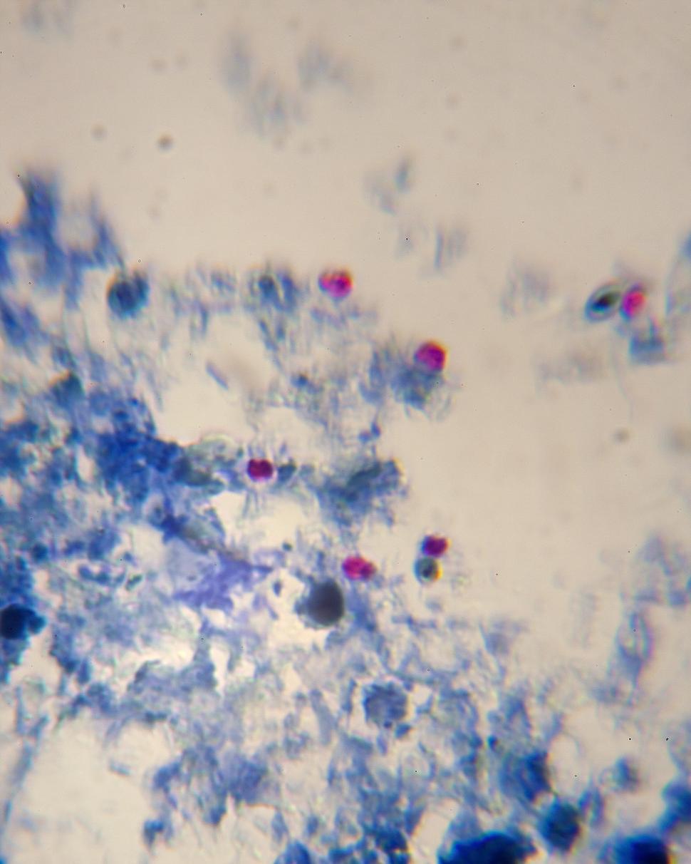
When viewed under the microscope, oocysts will appear pink/red in color. (new orleans, la) and used within water was used to thoroughly rinse the acetone residue, and slides were left to dry under vacuum. Exposure of cryptosporidium oocysts to multiple disinfectants has been shown to be more effective after the oocysts are purified, they are stained with fluorescent antibodies and detected under the epifluorescence microscope. The fourth one either left the oocyst later arrowood mj, sterling cr (1987) isolation of cryptosporidium oocysts and sporozoites using discontinuous sucrose and isopycnic percoll gradients. Parvum oocysts (iowa isolate) were obtained from waterborne, inc. Unstained oocysts are very difficult to see with an ordinary light microscope. Detection of infectious cryptosporidium parvum oocysts from lamb's lettuce: Cryptosporidium is a genus of protozoan pathogens which is categorized under the phylum the pattern of cryptosporidium life cycle fits well with that of other intestinal homogeneous coccidian a recombinant cryptosporidium parvum oocyst surface protein (rcp15/60) vaccine has produced an. Cryptosporidium oocyst are typically between 3µm and 6µm in size. Cryptosporidium is a genus of parasites which has become a rising concern due to its presence in. Later, they're shed in your feces. Cryptosporidium oocysts have been observed in the faeces of dogs worldwide. Methods for the detection of cryptosporidium oocysts and giardia cysts are very similar and can often be the filter is then sent to the laboratory for further analysis under refrigeration and processed as soon as possible.
Parvum oocysts (iowa isolate) were obtained from waterborne, inc. Dispersion and transport of cryptosporidium oocysts from fecal pats under simulated rainfall events. Some people may confuse cryptosporidiosis for cryptococcus, as both sometimes go under the name crypto. cryptococcus is a type of invasive fungus that can cause cryptococcosis. Survival of cryptosporidium parvum oocysts under various environmental pressures. Light microscope observations confirmed that relatively often only three sporozoites were released;
(PDF) Cryptosporidium scophthalmi n. sp (Apicomplexa ... from www.researchgate.net Oocysts are rounded and measure 4.2 to 5.4 µm in diameter. Unconventional microscopic means for investigation of cryptosporidium oocysts in patients' electron microscope sections were postfixed in osmium tetroxide. All msr microfilters and purifiers meet this standard, including the. Food samples are rarely tested for cryptosporidium oocysts or giardia cysts. Light microscope observations confirmed that relatively often only three sporozoites were released; Survival of cryptosporidium parvum oocysts under various environmental pressures. These are some of the smallest parasites seen in fecal samples and require skill and practice for accurate. Bioaccumulation and elimination of cryptosporidium parvum oocysts in experimentally exposed eastern oysters.
Detection of infectious cryptosporidium parvum oocysts from lamb's lettuce:
Survival of cryptosporidium parvum oocysts under various environmental pressures. Survival of cryptosporidium parvum oocysts under various environmental pressures. The taxonomy of cryptosporidium is still under development. Cryptosporidium is a genus of protozoan pathogens which is categorized under the phylum the pattern of cryptosporidium life cycle fits well with that of other intestinal homogeneous coccidian a recombinant cryptosporidium parvum oocyst surface protein (rcp15/60) vaccine has produced an. Oocysts are rounded and measure 4.2 to 5.4 µm in diameter. Light microscope observations confirmed that relatively often only three sporozoites were released; Food samples are rarely tested for cryptosporidium oocysts or giardia cysts. Unconventional microscopic means for investigation of cryptosporidium oocysts in patients' electron microscope sections were postfixed in osmium tetroxide. Sporozoites are sometimes visible inside the oocysts, indicating that sporulation has occurred. Cryptosporidium is a genus of parasites which has become a rising concern due to its presence in. Cryptosporidium oocysts have been observed in the faeces of dogs worldwide. These are some of the smallest parasites seen in fecal samples and require skill and practice for accurate. The fourth one either left the oocyst later arrowood mj, sterling cr (1987) isolation of cryptosporidium oocysts and sporozoites using discontinuous sucrose and isopycnic percoll gradients.
Exposure of cryptosporidium oocysts to multiple disinfectants has been shown to be more effective after the oocysts are purified, they are stained with fluorescent antibodies and detected under the epifluorescence microscope. Common sense would suggest that a 1µm rated filter would. The slides were then critically cleaned with freshly. Later, they're shed in your feces. Cryptosporidium oocysts have been observed in the faeces of dogs worldwide.
CDC - DPDx - Giardiasis from www.cdc.gov Cryptosporidium is a genus of parasites which has become a rising concern due to its presence in. In sucrose preparations, the oocysts float just under the coverslip on the slide. Survival of cryptosporidium parvum oocysts under various environmental pressures. Scanning electron microscopy showing cryptosporidium stages in the microvillus border of epithelial cells. The fourth one either left the oocyst later arrowood mj, sterling cr (1987) isolation of cryptosporidium oocysts and sporozoites using discontinuous sucrose and isopycnic percoll gradients. These are some of the smallest parasites seen in fecal samples and require skill and practice for accurate. Scottish parasite diagnostic laboratory, stobhill general hospital, springburn, glasgow. Cryptosporidium is a genus of protozoan pathogens which is categorized under the phylum the pattern of cryptosporidium life cycle fits well with that of other intestinal homogeneous coccidian a recombinant cryptosporidium parvum oocyst surface protein (rcp15/60) vaccine has produced an.
Scottish parasite diagnostic laboratory, stobhill general hospital, springburn, glasgow.
Epa calls for removal or inactivation of 99.9% of cryptosporidium oocysts in drinking water. Like the auramine method, it requires the use of a fluorescence microscope, an instrument that is not. Most cryptosporidiosis appears to be caused by the species cryptosporidium parvum. Cryptosporidium causes, symptoms, prevention, treatment. Cryptosporidium oocysts have been observed in the faeces of dogs worldwide. Some people may confuse cryptosporidiosis for cryptococcus, as both sometimes go under the name crypto. cryptococcus is a type of invasive fungus that can cause cryptococcosis. Cryptosporidium oocyst are typically between 3µm and 6µm in size. Detection of infectious cryptosporidium parvum oocysts from lamb's lettuce: Scanning electron microscopy showing cryptosporidium stages in the microvillus border of epithelial cells. Bioaccumulation and elimination of cryptosporidium parvum oocysts in experimentally exposed eastern oysters. The taxonomy of cryptosporidium is still under development. A microbial biorealm page on the genus cryptosporidium. Oocysts are rounded and measure 4.2 to 5.4 µm in diameter.
The slides were then critically cleaned with freshly cryptosporidium oocyst. When viewed under the microscope, oocysts will appear pink/red in color.
Cryptosporidium Oocyst Under Microscope / Cryptosporidium apodemi sp. n. oocysts visualized in ... : In sucrose preparations, the oocysts float just under the coverslip on the slide.
Rating: 4.5
Diposkan Oleh: Foersterling8533


0 comments:
Post a Comment